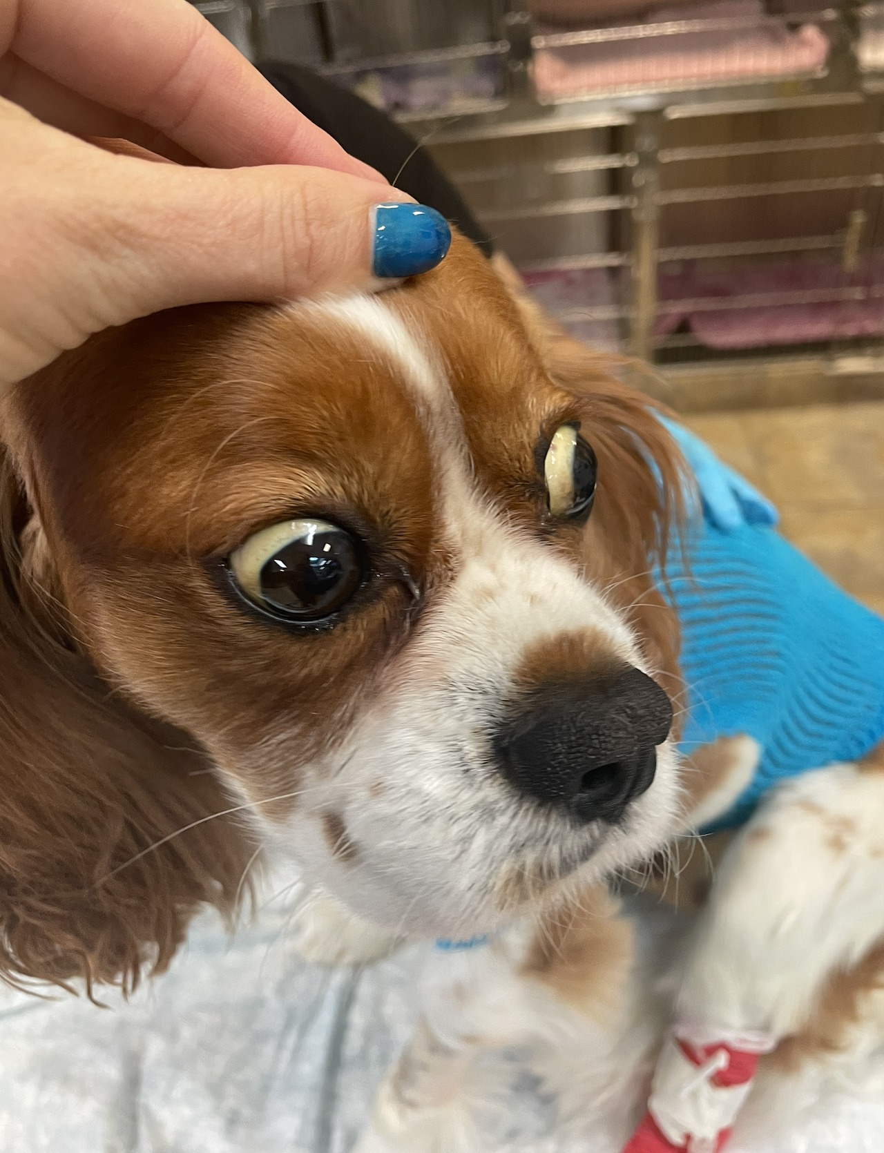
IMHA in Dogs and Cats: Diagnostics & Therapeutics
Immune-mediated hemolytic anemia (IMHA) is one of the most common—and most challenging—immune-mediated diseases seen in dogs, with cats affected less often. It is a life-threatening autoimmune condition where the immune system targets and destroys the body’s own red blood cells, leading to severe anemia and systemic complications. For veterinarians, early recognition, rapid stabilization, and systematic long-term management are critical to improving survival.
Pathophysiology & Clinical Picture
In IMHA, antibodies bind to red blood cell (RBC) surface antigens. This immune reaction leads to hemolysis through two key mechanisms:
Extravascular hemolysis (most common): Antibody-coated RBCs are removed by macrophages in the spleen and liver, producing spherocytes and often splenomegaly with hyperbilirubinemia.
Intravascular hemolysis (severe cases): Complement-mediated RBC lysis occurs in circulation, causing hemoglobinemia, hemoglobinuria, and sometimes acute kidney injury.
IMHA is classified as primary (idiopathic)—a true autoimmune disease without an identifiable trigger—or secondary, associated with infections (Babesia, Ehrlichia, Mycoplasma), neoplasia, medications, or other immune-mediated conditions.
Typical clinical signs reflect both anemia and hemolysis: pale mucous membranes, tachycardia, weakness, exercise intolerance, jaundice, dark urine, splenomegaly, and occasionally hepatomegaly. Fever, collapse, vomiting, and abdominal distension may also occur in severe cases.
Diagnostic Approach
Diagnosis requires rapid assessment and systematic exclusion of secondary causes.
Packed cell volume (PCV): Often <20%; plasma may be icteric or hemolyzed.
Blood smear: The hallmark findings are spherocytes, autoagglutination, polychromasia, and sometimes ghost cells.
CBC: Usually severe regenerative anemia, leukocytosis from inflammation, and occasionally concurrent thrombocytopenia (Evans syndrome).
Chemistry panel: Elevated bilirubin, possible liver enzyme changes, and renal parameters to assess for injury.
Coombs (Direct Antiglobulin) test: The gold standard, though false negatives are possible.
Saline agglutination: A quick, in-clinic supportive test.
Flow cytometry: Increasingly recognized as more sensitive, but not widely available.
Secondary causes must be excluded with infectious disease panels, FeLV/FIV testing in cats, and thoracic/abdominal imaging to rule out neoplasia or other triggers. Crossmatching and blood typing are essential prior to transfusion.
Case Highlight: Primary IMHA in a Labrador Retriever
A 7-year-old spayed female Labrador presented with lethargy, tachypnea, and icterus. PCV was 12%, blood smear revealed numerous spherocytes with autoagglutination, and a Coombs test was positive. She required transfusion and aggressive immunosuppression, stabilizing at a PCV of 25% after 10 days. This is a textbook case of primary IMHA requiring rapid intervention.
Therapeutic Management
Emergency Stabilization
The first priority is evaluating transfusion need. Packed red blood cells are the preferred product, targeting a PCV of 20–25%. Crossmatching is essential, and patients should be monitored closely during transfusion for reactions.
Primary Immunosuppressive Therapy
Prednisolone/Prednisone (2–4 mg/kg PO q12h): Remains the cornerstone, suppressing antibody production and reducing splenic sequestration.
Dexamethasone (0.5–1 mg/kg IV q24h): Used in severe, unstable patients until oral therapy is possible.
Improvement is expected within 24–48 hours (cessation of hemolysis), with reticulocytosis peaking in 3–5 days and PCV recovery within 1–2 weeks.
Second-Line Immunosuppressants
For refractory cases or to reduce steroid burden:
Azathioprine – delayed onset, requires careful monitoring for hepatotoxicity and myelosuppression.
Cyclosporine – faster onset (2–3 weeks), effective and steroid-sparing.
Mycophenolate mofetil – emerging option for difficult cases.
Rescue Options
IV immunoglobulin (IVIG): Can provide rapid, though temporary, benefit in severe cases.
Splenectomy: Considered when medical management fails or relapses are frequent.
Plasmapheresis: Reserved for specialized centers and critical refractory cases.
Complications and Concurrent Conditions
One of the most dangerous sequelae is thromboembolism, affecting up to 30% of IMHA patients. Preventive strategies include aspirin, clopidogrel, or heparin in high-risk patients.
Evans syndrome (IMHA + ITP) occurs in up to 15% of cases, carries a poorer prognosis, and demands aggressive multimodal immunosuppression.
Acute kidney injury, often secondary to hemoglobinuria, necessitates vigilant renal monitoring and fluid support.
Case Highlight: Secondary IMHA with Babesiosis
A 3-year-old Rhodesian Ridgeback presented with acute weakness and hemoglobinuria following a camping trip. PCV was 8%, blood smear confirmed Babesia canis, and Coombs test was positive. Treatment included blood transfusion, imidocarb therapy, and prednisolone, with recovery achieved over three weeks.
👉 Always investigate travel and exposure history to rule out secondary infectious causes.
Monitoring & Long-Term Care
During the acute phase, hospitalized patients require frequent PCV checks, vitals monitoring, and serial chemistries. Once stable, long-term therapy involves a gradual steroid taper once PCV remains >30% and hemolysis subsides.
Relapse occurs in 15–20% of cases, often from premature tapering or concurrent stress/illness. Maintenance immunosuppression is sometimes required, particularly in cases of Evans syndrome.
Prognosis
Primary IMHA: 60–70% survival to discharge, with 50–60% achieving long-term survival.
Secondary IMHA: Prognosis varies widely depending on the underlying cause.
Evans syndrome: Guarded, with survival rates of 40–50%.
Thromboembolism: Worsens prognosis significantly.
Favorable prognostic factors include younger age, higher initial PCV, gradual onset, and rapid response to therapy. Poor prognostic indicators include intravascular hemolysis, concurrent thrombocytopenia, and thromboembolic events.
Client Education & Prevention
Owner education is essential for success:
Medication compliance prevents relapse.
Monitoring for steroid side effects (PU/PD, panting, weight gain) is crucial.
Recognizing relapse signs such as pale gums, jaundice, or lethargy ensures rapid intervention.
Risk reduction strategies include careful vaccination decisions in predisposed breeds, vector control for blood parasites, and thoughtful drug choices. Long-term follow-up and tailored vaccination/breeding recommendations may also be indicated.
Key Takeaways
IMHA is one of the most challenging immune-mediated diseases in veterinary medicine.
Rapid recognition, transfusion support, and immunosuppressive therapy are lifesaving.
Rule out secondary causes such as infectious disease or neoplasia.
Long-term management and client compliance are essential, as relapse and complications are common.
📚 References:
Garden OA, Kidd L, Mexas AM, et al. ACVIM consensus statement: diagnosis of IMHA in dogs and cats. JVIM. 2019.
Swann JW, Garden OA, Fellman CL, et al. ACVIM consensus statement: treatment of IMHA in dogs. JVIM. 2019.
Kohn B, Weingart C, et al. IMHA in cats: outcomes and therapy. JVIM. 2006.
Weinkle TK, Center SA, et al. Prognostic factors in canine IMHA. JAVMA. 2005.
Barton JC, German AJ, et al. Proteinuria in dogs with immune-mediated disease. JVIM. 2025.
Nivy R, Sutton G, Bruchim Y. Carboxyhemoglobin as a biomarker in hemolytic anemias. Vet Clin Pathol. 2018.

