AI Lymphoma Detection
Faster, confident diagnostics means our patients get faster access to life saving treatments.
Fast, objective lymphoma detection powered by AI
Pathview brings rapid, AI-powered cytological assessment directly to your clinic for confident large cell lymphoma diagnosis in dogs and cats
Detection accuracy in current validation
Availability for rapid diagnosis
Cells analyzed per sample
Built for modern veterinary practice
We're bringing specialist-level analysis directly to your clinic with AI-powered technology that enhances efficiency and confidence
Rapid turnaround
Get results while your patient is still in the clinic. Make immediate treatment decisions without waiting days for specialist review.
Pathologist confirmation
Every result is reviewed by our board-certified veterinary clinical pathologists for complete diagnostic confidence.
Thorough analysis
The AI analyzes thousands of cells across multiple high-powered fields—far more than manual counting allows.
Consistent quality
Standardized assessment reduces inter-observer variability. Get the same reliable results regardless of when you submit.

How the AI pipeline works
Our three-stage AI pipeline mimics the workflow of an experienced clinical pathologist
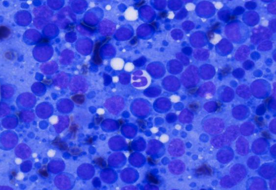
Assessment
The AI scans the entire slide to identify diagnostic quality regions with optimal cell distribution and staining. Poor quality areas are automatically filtered out to ensure reliable analysis.
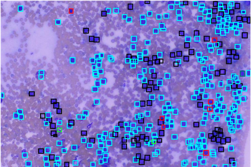
Classification
In high-quality regions, the AI meticulously detects and catalogs every relevant cell—small and large lymphocytes, lysed cells, other white blood cells, and mitotic figures.
Analysis
The system analyzes cell populations, calculating total cell counts, large-to-small lymphocyte percentages, and intact-to-lysed cell ratios to generate a clear diagnostic classification.
Validated accuracy you can trust
Current detection accuracy for large cell lymphoma
Trained on thousands of manually labeled cells from diverse cases, our AI demonstrates excellent agreement with board-certified veterinary clinical pathologists.
AI-enhanced vs. traditional cytology
Cytology alone
- Subjective cell population estimates
- Inter-observer variability in interpretation
- Limited cell counting in practice
- Requires specialist review for confidence
- Turnaround time: days to weeks
AI-powered assessment
- Objective, quantitative cell counts
- Standardized, reproducible results
- Thousands of cells analyzed per sample
- Pathologist confirmation included
- Turnaround time: same day, often within hours
Test specifications
Everything you need to know about submitting samples and interpreting results
Validated for both canine and feline lymph node aspirates
Single slide submission with standard staining
Optimized for high-grade lymphomas with large immature lymphocytes
"Images consistent with large cell lymphoma" or "Images not consistent with large cell lymphoma"
Ready to enhance your lymphoma diagnostics?
We're here to support your clinic's diagnostic needs every step of the way. Get started with Pathview AI Lymphoma Cytology or schedule a demo to see it in action.